SDS-PAGE Technique
SDS-PAGE, which full name is sodium dodecyl sulfate (SDS) polyacrylamide gel electrophoresis, is a powerful tool for protein analysis (molecular masses between 5 and 250 kDa) due to its availability and reproducibility. The combination of SDS and polyacrylamide gel allows this technique to eliminate the influence of proteins' structure and charge, and achieve separation only based on differences in their molecular weight. Moreover, SDS-PAGE can also be used for purity characterization of PEGylated proteins after purification steps and quantification of PEGylation extent by studying the molecular weight of appeared bands on gel.
Principles
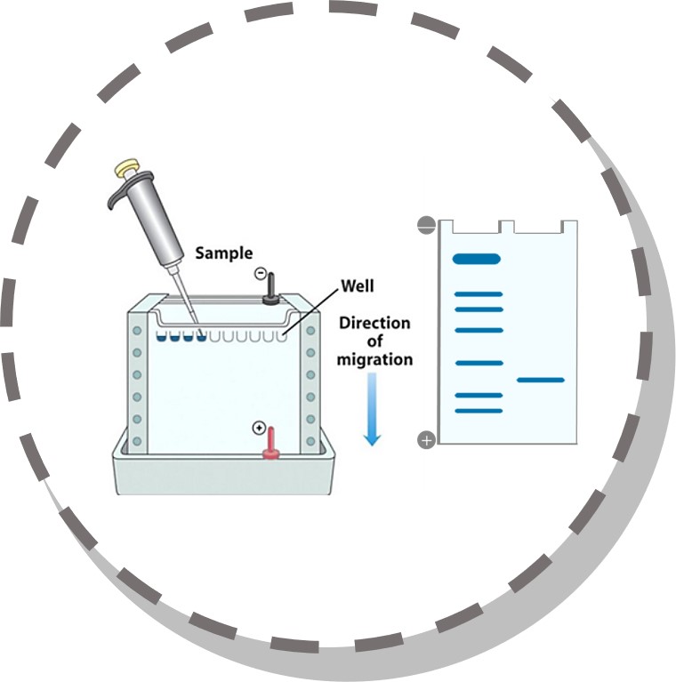
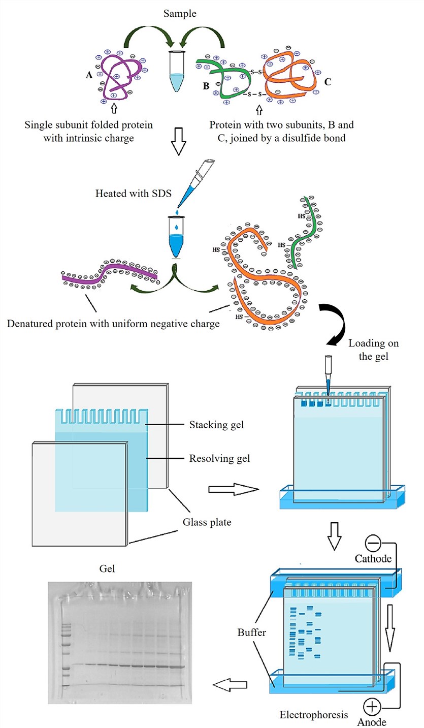 Fig. 1 Schematic illustration of polyacrylamide gel electrophoresis. (Academic Press, 2018: 317-351)
Sodium dodecyl sulfate, namely SDS, is an anionic detergent which can break the intra- and intermolecular hydrogen bonds, further destroying the secondary and tertiary structure of protein molecules. Moreover, strong reducing agents, such as mercaptoethanol and dithiothreitol, can break the disulfide bond between cysteine residues. In SDS-PAGE, due to the addition of SDS and reducing agent, the molecules are depolymerized into polypeptide chains and amino acid side chains, which are then combined with SDS combine to form protein-SDS micelles. Therefore, the biological activity of the protein is lost or weakened during electrophoresis, but its activity can be restored or partially recovered after electrophoresis.
When the ionic strength is low, SDS, which is mainly in the form of monomers, can bind to protein to form a protein-SDS complex. Because there are large numbers of negatively charged groups on SDS, which makes the difference in charge between different proteins masked. The shape of the SDS-protein complexes are all elliptical rods, and the length of the rods is related to the molecular weight of the protein subunits. Therefore, in SDS-PAGE, the mobility of protein is only related to molecular weight, which can be used to separate different proteins.
What is 2D SDS-PAGE?
With the advancement of science and technology, researchers have developed a more efficient electrophoresis technology, two-dimensional SDS-PAGE (2D SDS-PAGE). This technique consists of two parts of electrophoresis. the first one is usually the isoelectric focusing (IEF) gel electrophoresis, in which denatured proteins are separated according to their isoelectric points (pI). The second part is SDS-polyacrylamide gel electrophoresis based on the molecular weight of protein. Using 2D SDS-PAGE can separate complex protein components on a two-dimensional plane.
IEF
The separation process of the protein mixture in the first dimension is based on the difference pf protein’s pI, which is called IEF. As well known, protein is an amphiphilic molecule, and its surface charge varies according to the pH of the environment. When the pH is higher than its pI, the protein displays negative charges, and vice versa. Under the action of the electric field, the protein molecules will drift towards the anode or the cathode, respectively. When reaching a pH position where the pI is same, the protein is not charged and no longer drifts. According this, when the voltage is applied, the positively charged molecules will move to the cathode, while the negatively charged molecules will move to the anode, thus forming a continuous pH gradient. As a result, when the protein migrates to the pH position where the pI is same, the charged state reaches equilibrium and no longer migrates, and the protein molecules are separated.
Different Methods for SDS-PAGE for PEGylated Proteins
- Different staining methods, such as barium iodide staining and Coomassie brilliant blue staining, can be utilized to determine the free and conjugated PEG molecules in gel and the protein in gel, respectively.
- SDS-PAGE can be used to compare the extent of attached PEG after solid phase and liquid phase PEGylation based on the number of appeared bonds on the gel.
- As the apparent hydrophobicity of a PEGylated protein increases with the number of attached PEG chains, duplicated SDS-PAGE (12.5% nonreducing) by Coomassie blue staining and PEG-staining can be used to detect the purified PEGylated species.
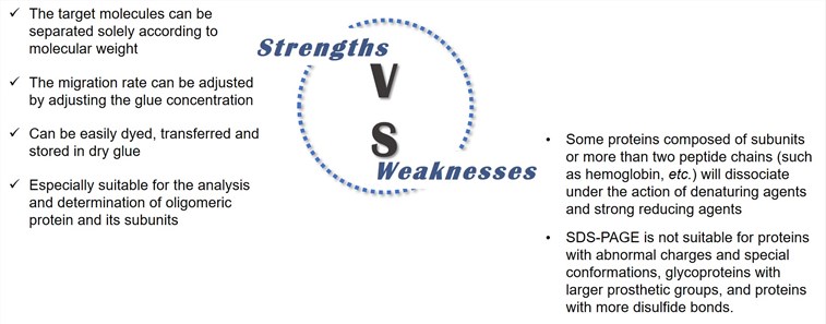
Strengths & Weaknesses of SDS-PAGE
Reference
- Büyükköroğlu G, Dora D D, et al. Techniques for protein analysis[M]//Omics Technologies and Bio-Engineering. Academic Press, 2018: 317-351.