
Halide (chloride, bromide) PEG
Reactive Intermediates, Synthesis
Article Body

Due to its good hydrophilic and compliant properties, polyethylene glycol can improve the pharmacokinetic and pharmacodynamic properties of drugs, and its modification on the surface of targeted nano preparation can increase the retention time and concentration of drugs in vivo. The physicochemical modification methods of polyethylene glycol-targeted nano-formulations include physical insertion and chemical bonding of polyethylene glycol lipid derivatives. Understanding the effects of PEG parameters (such as PEG relative molecular mass, modification density, and spatial conformation) on the performance of targeted nanomaterials can provide a reference for better construction of PEG-targeted nanomaterials.
Researchers prefer to insert the PEG-lipid with the characteristics of a low price, wide varieties, easy synthesis, high safety, good biocompatibility, and better modification effect into the nanomaterials physically. As a result, the steric hindrance effect prevents the aggregation of nanoparticles, reduces the recognition and adsorption of plasma protein, prolongs the retention time in the blood, and increases the stability of the nanomaterials.
In a research group, Zhang et al. prepared dual-targeted nanoparticles using DSPE-PEG anisamide modified rHDL/(DCA-PEI/p53) complexes. Through the dual mediation of Sigma and SR-BI receptors, they also delivered sodium dichloroacetate (DCA) and the therapy gene p53 into the tumor cells. The nanoparticle has uniform particle size, neutral surface charge, and little toxicity to normal cells. Therefore, through the synergistic action of DCA and p53, it effectively prevents the growth of tumor cells and reduces the tumor volume of mice to (443.23±78.13) mm3.
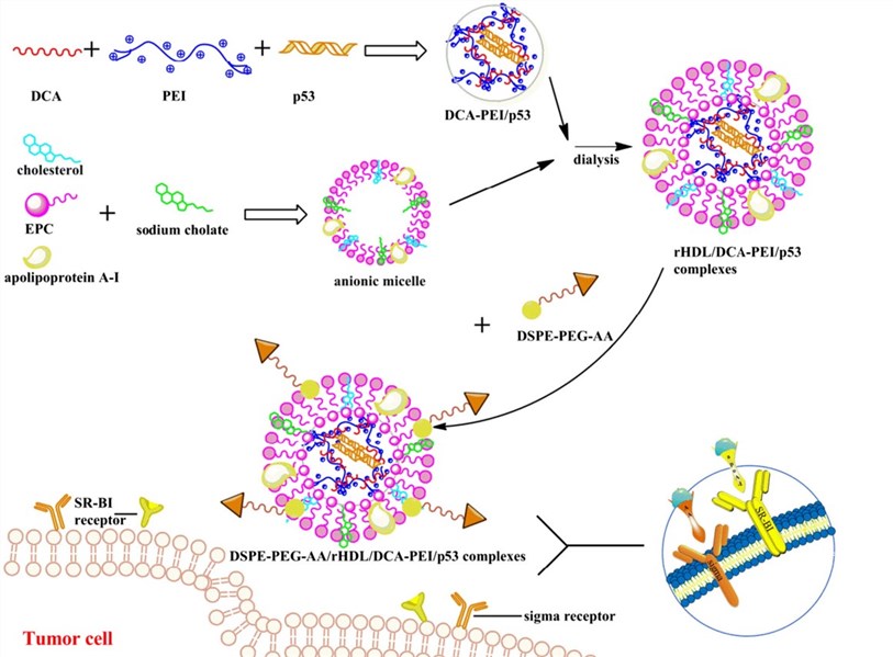 Fig. 1. Research applications of DSPE-PEG (Mater Sci Eng C. 2016, 64(1): 208-218).
Xu et al. made vesicles or liposomes with cholesterol succinic acid monoester and mPEG-cholesterol derivative or mPEG-DSPE. They wrapped calcein and then investigated the pharmacokinetics. The results showed that the plasma clearance rate of calcein in mPEG-DSPE modified liposomes was significantly increased, and the phenomenon of accelerated blood clearance was obvious. The plasma clearance rate of mPEG-cholesterol derivative-modified liposomes was almost unchanged, suggesting that these liposomes could effectively slow down or eliminate the accelerated blood clearance phenomenon.
Yameaqo et al. made PEG-fatty acid esters and decyl-grafted cyclodextrin derivatives (CD-C10) through nano co-deposition to produce PEG-surface modified nanoparticles. The in vitro drug release time is 96h, and the fatality rate of half is 13nmol/L, which is much better than the nanoparticles without PEG-surface modification.
PEG itself contains only -OH but it can be attached different reaction groups by chemical reactions, and then modify the nanoparticle surface.
PEG terminal group can be chemically modified to -NH2 and PEG can be chemically bonded to nanoparticles through dehydration condensation with -COOH. Chen et al. reacted long-chain oleoyl fatty acids with mPEG-NH2 and formed micelles by self-assembly, and finally prepared PEG-coated superparamagnetic iron oxide nanoparticles. The PEG layer on the surface of the nanoparticles prevents the settlement of the nanoparticles, which makes them highly stable in pH3-10 aqueous solution or 0.3mol/L NaCl salt solution.
Zhou et al. connected mPEG-COOH with branched PEI through an amide bond, and then synthesized nanoparticles (Au PENPs) with PEG-modified PEI. Due to the low cytotoxicity and the hemolysis, Au PENPs can be used in CT imaging. PEG modification greatly improves the biological compatibility of AuNPs, prolongs the half-life from 11.2h to 31.76h, reduces the absorption of macrophages, and they can be more widely used in tumor CT imaging.
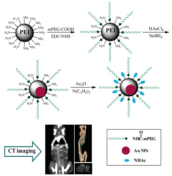 Fig. 2. Schematic illustration of the preparation of pegylated Au PENPs for CT imaging applications (ACS Appl Mater Interfaces. 2014, 6(19): 17190-17199).
The PEG-modified drug delivery system can prolong its circulation time in the body but requires rapid drug release after reaching the targeted site to achieve a therapeutic effect. Hou et al. oxidized PEG-OH to CHO-PEG-CHO, which was chemically modified on Fe3O4 nanoparticles coated with polydopamine (PDA-coated Fe3O4NPs). The PEG chain extends into the solution and forms a brush-like structure under certain conditions, which improves the hydrophobicity and rigidity of the magnetic nanoparticles. PEG acts as the link between the enzyme and its support, which makes PDA magnetic materials more suitable for enzyme immobilization.
Qian et al. have replaced the CTAB coated with gold nanoparticles with PEG-SH and dithioth reitol (DTTC) to form PEG and dye-coated gold nanorods (PEG-DTTC-GNRS). Since they are not toxic to tissues, organs, and nerves, when the maximum dose is 8μg/mL, the cell survival rate is still up to 86%. This method could be used for in vivo sentinel lymph node (SLN) imaging, tumor targeting and diagnosis, and provides a new path for achieving an efficient and non-toxic optical imaging method.
By linking PEG-NHS with dendritic macromolecules containing symmetric analogs of L-lysine or lysine, Kaminskas et al. made PEG-modified poly-lysine dendritic macromolecules containing doxorubicin. This preparation has the characteristics of pH-sensitive doxorubicin release, long blood circulation time, and tumor targeting.
Feng et al. connected PLA with the hydroxyl group of PEG through both ends of the ester bond, synthesized HO-PLA-PEG-PLA-OH, connected pyropheophorbide a (PPa) and F3 peptide, then prepared nanoparticles of co-transfer photosensitized PPa and chemotherapy drug Paxil, and finally prepared nanoparticles of PPNP. In this nanoparticle, the encapsulation rate of the two drugs is high, (71.07 ± 2.57) % and (67 ± 3.05) % respectively. It actively targets the tumor, realizing the synergistic effect of chemotherapy and photodynamic therapy.
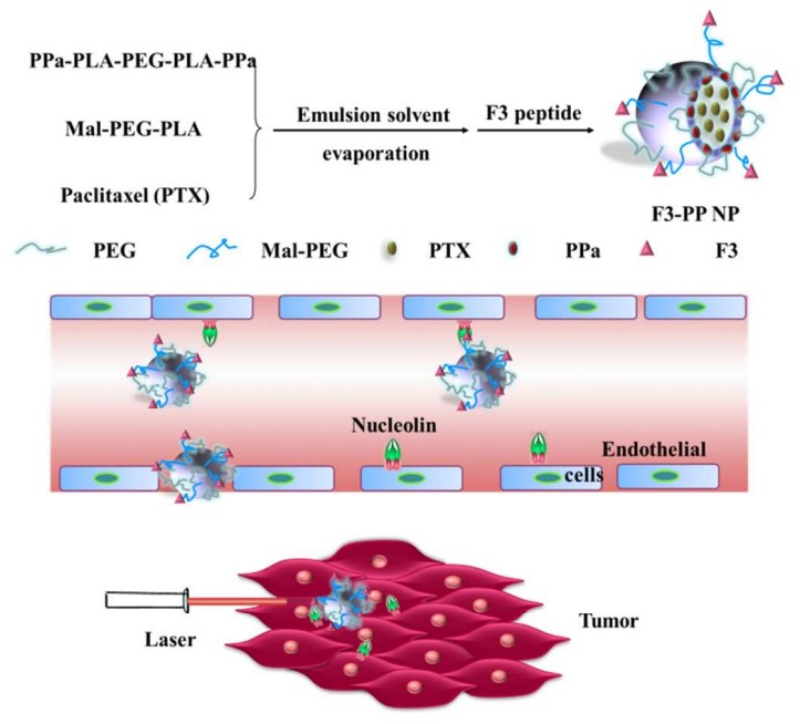 Fig. 3. Research applications of PEG-PLA nanoparticles in cancer therapy (ACS. 2016, 8(28): 17817-17832).
Since PEG is hydrophilic, free of charge, and easy to be modified onto the surface of nanoparticles, most researchers have regarded PEG-modified nanoparticles as the best choice for long-cycle nanoparticles. However, PEG's ability to repel proteins and macrophages depends on different parameters, such as relative molecular mass, density, spatial conformation, and flexibility.
At present, there are not many applicable methods for the modification of PEG onto the surface of nanoparticles and the existing techniques are still flawed. Therefore, more efforts should be made to improve the PEG modification techniques. And the combination of PEG modification methods with analytical methods can also help confirm the modification results. BOC Sciences provides PEGylation services for various types of nanoparticles, such as liposomes, dendrimers, and polymeric nanoparticles. We use a variety of PEGylation techniques, including covalent conjugation, non-covalent adsorption, and encapsulation. BOC Sciences can modify the size, shape, and surface chemistry of nanoparticles to optimize their performance in drug delivery applications.
References
Planning to purchase or looking for custom solutions?
Get in touch with our experts today! We'll respond within 24 hours to discuss your project needs and provide a customized quote.
From custom PEG synthesis to PEGylation support, explore tailored services designed to meet the specific needs of your research.
Browse related content to gain a deeper understanding of PEG synthesis and manufacturing.

Halide (chloride, bromide) PEG
Reactive Intermediates, Synthesis

Gene Delivery, Cationic Carriers

Drug Conjugates, Delivery

Drug Delivery, Material Design

Liposomes, Drug Delivery

Sustained Release, Implants

Crosslinking, Protein Conjugation

Crosslinking, Surface Coating

Click Chemistry, Hydrogels

Amine Coupling, Conjugation
Online Inquiry
Case Studies and Success Stories