PEG for Imaging Diagnosis
Accurate diagnosis of cancer is essential for improving the survival rate of cancer patients. With advances in the synthesis of a variety of nanomaterials, there has been a surge of interest in the use of nanoparticles as imaging probes or contrast agents for clinical techniques. However, because of the characteristic properties of traditional tools do not always meet the need of personalized cancer diagnosis, PEGylated nanoparticles have also been explored in diagnostics in which it has found its role in tracking agents and/or contrast agents used in imaging techniques, such as computed X-ray tomography (CT), magnetic resonance imaging (MRI), and ultra-sonography.
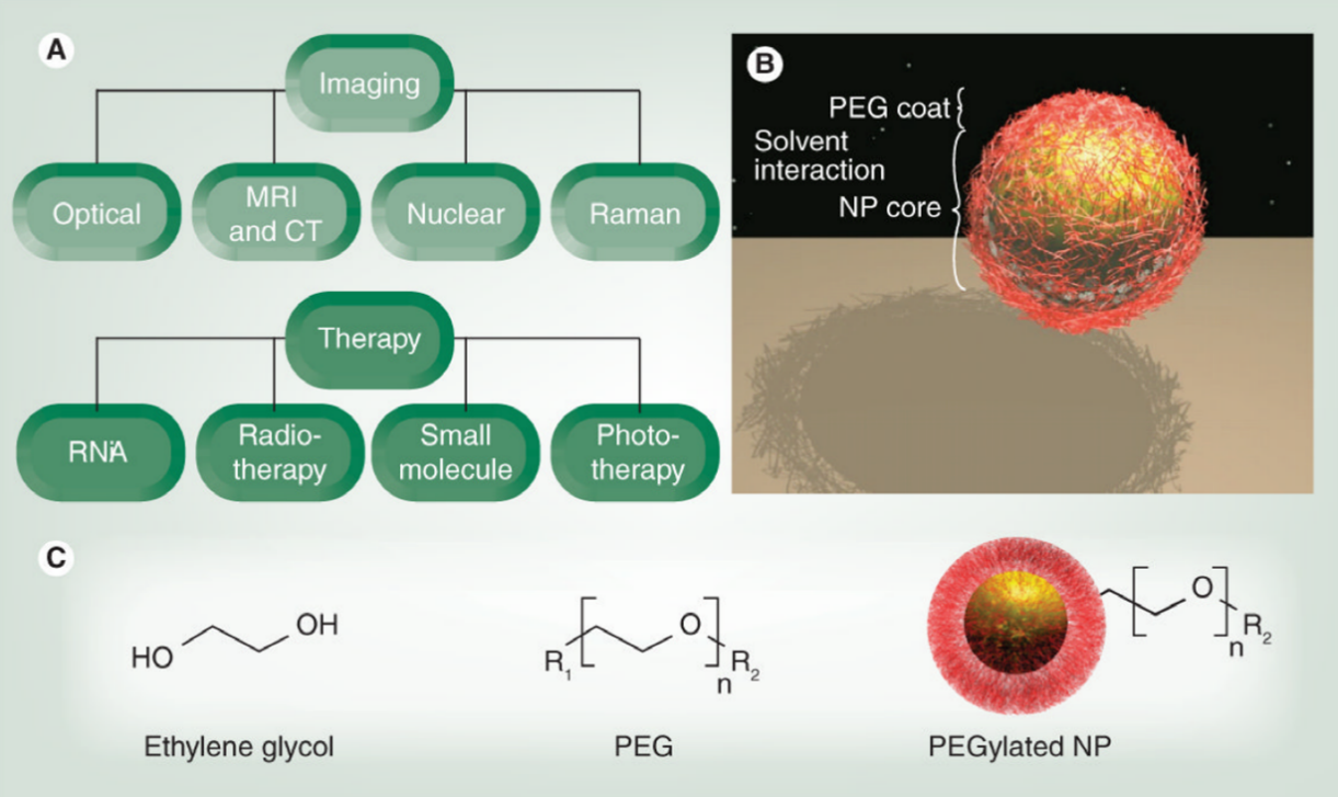 Fig. 1 Schematic illustration of PEGylated nanoparticles act as imaging tools. (Nanomedicine 2011, 6 (4), 715-728.)
Background
Typical examples include magnetic nanoparticles (e.g., paramagnetic gadolinium (Gd3+) chelates) as magnetic resonance contrast agents, quantum dots and gold nanoparticles (GNPs) as optical imaging probes, as well as other radioactive materials used in CT and ultrasound. However, those traditional nanoparticles are disadvantaged by impaired target specificity and inadequate information on the lesion location in the case of cancer. For example, they can be uptake by the reticuloendothelial system (RES), in which nanoparticles are rapidly shuttled out of circulation to the liver, spleen or bone marrow, and nonspecific bound to non-targeted or non-diseased areas. Concerns about nanoparticles' toxicity often arise because of this RES accumulation.
The addition of polyethylene glycol (PEG) to the nanoparticle surface (i.e., PEGylation) can notably reduce many of these challenges. PEG is a coiled polymer of repeating ethylene ether units with dynamic conformations. In both imaging and drug delivery applications, chemical modification using PEG polymers can reduce RES uptake and prolong the circulation time. Owing to the passivated surfaces, the aggregation and nontargeted association phenomena are diminished, resulting in so-called `stealth' behavior. Moreover, the enhanced permeability and retention (EPR) effect can be modulated due to nanoparticle size changes via addition of PEG polymers. Due to these attributes, PEGylated nanoparticles generally accumulate in the liver a half to a third of the amount of non-PEGylated nanoparticles and demonstrate higher tumor accumulation versus background.
Fluorescence Imaging
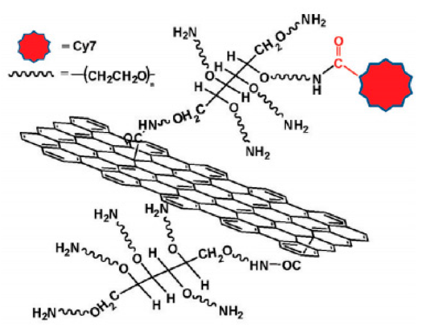 Fig. 2 Schematic illustration of graphene oxide modified with multiple-arm PEG chains for fluorescence imaging. (Pharmaceutics 2019, 11 (7), 327)
The activation of graphene oxide (GO) surfaces occurs by nucleophilic substitution reaction of the hydroxyl groups on the grapheme oxide with chloroacetic acid in the presence of NaOH to convert the -OH groups to a more reactive -COOH group (GO-CH2-COOH). Then, six arm amine-PEG is conjugated with GO-CH2-COOH through the activation of the carboxylic acid moiety with EDC and the formation of an amide bond. The remaining amines of the PEG are used for further attachment such as an infrared labeling molecule Cy7 through amide bonds.
Magnetic resonance Imaging
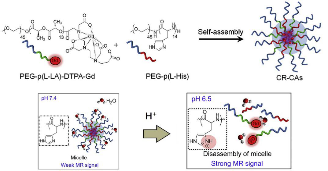 Fig. 3 Schematic representation of the self-assembled micelle used for MRI contrast agents. (Biomaterials 2014, 35 (1), 337-343)
Magnetic resonance imaging (MRI) with a contrast agent has been widely used as a powerful tool for cancer diagnosis. The most extensively and clinically used MRI contrast agents are paramagnetic gadolinium (Gd3+) chelates. However, the Gd3+-based contrast agents is compromised by their intrinsic low efficiency, which results in a need for high doses. Furthermore, most MRI contrast agents are nontargeted for cancer and passively distributed throughout the body. Incorporation of PEG in such systems has shown to further enhance their efficiency through site selective biodistribution.
X-ray Computed Tomography Imaging
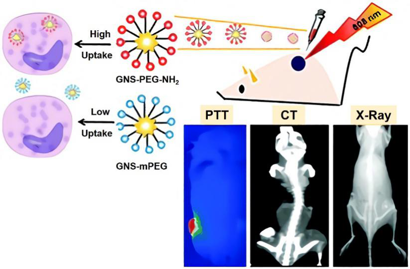 Fig. 4 Preparation of gold nano-stars functionalized with amine-terminated PEG for X-ray/CT imaging. (Journal of Materials Chemistry B 2015, 3 (21), 4330-4337)
X-ray computed tomography (CT) is one of the most useful diagnostic tools in hospitals. Current contrast agents for CT are based on iodinated small molecules because, among nonmetal atoms, iodine has a high X-ray absorption coefficient. Iodinated compounds, however, allow only very short imaging times due to rapid clearance by the kidney, which can also cause them to have renal toxicity. Currently, researchers have reported a biocompatible PEG polymer coated gold nanoparticles as a potential CT contrast agent. Due to the anti-biological fouling property, PEG can be modified onto the gold nanoparticles' surfaces, preventing from rapid clearance from the bloodstream due to uptake by the reticular endothelial system, including macrophages in the liver and spleen.
Ultrasound Molecular Imaging
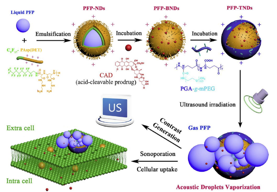 Fig. 5 Fabrication of functional nanodroplets coated with PGA-g-mPEG and the mechanisms of ultrasound-triggered imaging (Colloids and Surfaces B: Biointerfaces 2019, 174, 416-425)
Ultrasound molecular imaging is achieved using targeted microbubble contrast agents, which are highly reflective ultrasound materials that can target specific tissue imaging using specific biomarkers. The imaging of targeted microbubble ultrasound contrast agents in thrombosis and inflammation has attracted increased research attention, and the targeted specificity and binding stability of the contrast agent are beneficial for early diagnosis. Researchers have functionalized nanodroplets with PEG chains to endow them with good sustainability, low cytotoxicity and good biocompatibility. In ultrasound imaging study, this kind of composite nanodroplets produced significant contrast with ultrasound irradiation (3.5 MHz, MI=0.08) at 37 ℃.
References
- Rahme, K.; Dagher, N., Chemistry routes for copolymer synthesis containing PEG for targeting, imaging, and drug delivery purposes. Pharmaceutics 2019, 11 (7), 327.
- Jokerst, J. V.; Lobovkina, T.; et al. Nanoparticle PEGylation for imaging and therapy. Nanomedicine 2011, 6 (4), 715-728.
- Tian, Y.; Luo, S.; et al. Wang, S., Gold nanostars functionalized with amine-terminated PEG for X-ray/CT imaging and photothermal therapy. Journal of Materials Chemistry B 2015, 3 (21), 4330-4337.
- Gao, J.; Yu, B.; et al. Ultrasound triggered phase-change nanodroplets for doxorubicin prodrug delivery and ultrasound diagnosis: An in vitro study. Colloids and Surfaces B: Biointerfaces 2019, 174, 416-425.
- Kim, K. S.; Park, W.; et al. A cancer-recognizable MRI contrast agents using pH-responsive polymeric micelle. Biomaterials 2014, 35 (1), 337-343.