How are Liposomes Different from Micelles?
What are Liposomes?
Liposome is an artificial carrier membrane. In the water, the hydrophilic head of the phospholipid molecule is inserted into the water, and the hydrophobic tail of the liposome extends to the air, and after agitation, a spherical liposome of bilayer lipid molecules is formed. This bilayer structure is the same as the skin cell membrane structure, which helps the skin to absorb drugs or products, that is, the principle of liposome in vitro application. Liposomes can be used for genetic modification, or for the preparation of drugs. Utilizing the feature that liposomes can fuse with cell membranes, drugs can be delivered into cells. The main forms of interaction between liposomes and cells include intermembrane transport (lipid exchange of cell membranes), contact drug release, adsorption, fusion and endocytosis, which are the principles of liposomes in vivo.
Preparation of Liposomes
Ethanol injection method, ultrasonic method, liposome extruder extrusion method, high pressure homogenization method, thin film dispersion method, reverse phase evaporation method, and freeze-thaw method are commonly used preparation methods for liposomes. The main preparation steps and the characteristics of the liposome after preparation are as follows, wherein ultrasonic method, high pressure homogenization method and freeze-thaw method can be combined with other methods for preparation.
| Preparation | Preparation Steps | Liposome Characteristics |
|---|---|---|
| Ethanol Injection Method | Dissolve the drug and embedding materials in butyl ethanol, inject it into ethanol in PBS solution at a certain temperature and at a certain speed at a certain speed, and evaporate the ethanol to dryness. | Prepare single-lamellar liposomes, a few are multi-lamellar liposomes, and the problem of ethanol residue is difficult to solve. |
| Ultrasound | The prepared liposomes are obtained after ultrasonic treatment by Comtech or probe. | To prepare single-chamber liposomes, the ultrasonic treatment of the probe is easy to cause damage to the liposomes due to excessive peripheral temperature. |
| Liposome Extruder Extrusion Method | The prepared liposomes were extruded through a liposome extruder. | Polycarbonate egg membranes with different pore sizes can be used to prepare single- or multi-lamellar liposomes with a uniform particle size. If there are too many impurities in the liposome formula that can easily block the polycarbonate egg membrane, it can be used together with high-pressure homogenization. |
| High Pressure Homogenization | The prepared liposomes were obtained after being homogenized by a high-pressure homogenizer. | The preparation is mainly based on single-chamber plastids, and at the same time, it can reduce the particle size to improve the encapsulation efficiency and stability. |
| Thin Film Dispersion Method | Membranes such as phospholipids and fat-soluble drugs are dissolved in an appropriate amount of chloroform or other organic solvents (water-soluble drugs can be directly added to the solution), and the rotary evaporation is performed until a mixed film of the membrane material and the drug is formed on the bottle wall. The solution without adding water-soluble drugs, and adding a small amount of glass beads and continuing to rotate until a uniform liposome solution is formed. The prepared liposomes can be reduced in particle size by subsequent treatment such as ultrasound. | Prepare multi-lamellar or large single-lamellar liposomes, and the main ones are single-lamellar liposomes after ultrasonication. |
| Reverse Phase Evaporation | Add a certain proportion of membrane materials into organic solvents (petroleum ether, chloroform) to dissolve. If the drug is fat-soluble, it is added to the organic solvent, and if the drug is water-soluble, it is added to the aqueous phase. After ultrasonic mixing, the organic solvent was removed by rotary evaporation to form liposomes. | Large unilamellar liposomes. |
| Freeze-Thaw Method | The prepared liposomes were obtained by repeated freezing and thawing. | Single-chamber or multi-chamber repeated freezing and thawing can improve the encapsulation efficiency. |
Structure of Liposomes
According to structural features, liposomes can be classified into unilamellar liposomes, multilamellar liposomes, and multilamellar liposomes. Unilamellar liposomes are liposomes composed of monolayer bilayers, which are divided into small unilamellar liposomes and large unilamellar liposomes, with particle sizes between 20-80nm and 100nm-1um. Multilamellar liposomes are vesicles with a particle size of 1-5 μm composed of multiple bilayers. Multivesicular liposomes refer to non-concentric vesicles, liposomes with a particle size of about 5-50um, which can encapsulate water-soluble drugs and have the characteristics of low leakage.
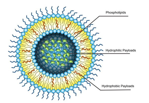 Fig. 1. Structural features of liposomes.
According to charge, liposomes can be divided into neutral liposomes, negatively charged liposomes and positively charged liposomes. Neutral liposomes are prepared from neutral phospholipid materials such as phosphatidylcholine choline. Negatively charged liposomes and positively charged liposomes are prepared by mixing a neutral phospholipid as an auxiliary material with a variety of negative or positive materials. Negative materials refer to phosphatidic acid, phosphatidylinositol, phosphatidylserine, and phosphatidylglycerides. Positive materials often include double-chain quaternary ammonium salt surfactants such as octadecylamine and dioctacyl-dimethylamine salt (DODAC). The charge of liposome directly affects its various properties, such as encapsulation efficiency, targeting, stability and so on. In addition, liposomes can also be divided into general liposomes and special performance liposomes according to their performance. Special performance liposomes refer to liposomes formed by changing the materials of liposomes, such as sensitive, heat-sensitive, magnetic and light-sensitive liposomes.
Characteristics of Liposomes
Targeting: The targeting of liposomes can be divided into passive targeting and active targeting. Passive targeting is greatly influenced by the particle size of liposomes. However, active targeting is more complicated and includes four types: chemical targeting, physical targeting, antibody targeting, and receptor targeting.
Sustained release: In the human body, the drug carried by the liposome is released slowly, which can greatly prolong the action time, delay renal excretion and metabolism, reduce toxicity, and avoid the inconvenience of needing to administer a small amount to the patient multiple times.
Compatibility of fat-soluble and water-soluble drugs: The phospholipid bilayer in the liposome wall material can carry fat-soluble drugs and the interior of the vesicle can carry water-soluble drugs, so it is a good biological carrier.
Non-toxic safety of liposomal membrane lipids: Studies have shown that even if the amount of lecithin used is large, it still has high safety. This component is also the main component of biological cell membranes.
Increase the stability of embedded drugs: Drugs (vaccine, insulin) that are easily decomposed and destroyed when entering the human body are prepared into liposomes to avoid damage to them by the internal environment and improve the stability of the ingredients.
What are Micelles?
In aqueous solution, when the surfactant reaches a certain concentration, the molecules self-assemble to form ordered thermodynamically stable colloidal aggregates. The mechanism of micelle formation is that after the surfactant with adsorption capacity reaches a saturated state, the excess surfactant is dispersed in the aqueous solution. Due to the presence of hydrophobic groups, the repulsion between water molecules and surfactants is stronger than the attraction. Hydrophobic groups associate under the action of van der Waals force to form the inner core of micelles, and hydrophilic groups face outward to form the outer layer of micelles, which are stably dispersed in aqueous solution.
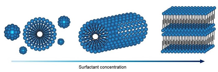 Fig. 2. Micellar structure evolution process.[1]
When the surfactant concentration was close to the critical micelle concentration (CMC), the micelles were spherical. When the concentration continues to increase, the number of associated molecules increases, and the micelles are in the form of cylinders or plates, and even complex forms such as stars and helices. The morphology of micelles is related to their free energy. Surfactants are amphiphilic molecules. The lipophilic tails gather inside the micelles because of their hydrophobicity, while the hydrophilic heads extend to the outside because of their polarity, and protect the hydrophobic groups inside the micelles. The main components of micelles are amphiphilic molecules, so in addition to being soluble in polar solvents (such as water), they are also soluble in non-polar solvents in the form of reverse micelles.
Characteristics of Micelles
Small particle size: The particle size of micelles is in the range of 10-100 nm, which is not easy to be recognized and captured by the reticuloendothelial system (RES) in the blood circulation system. The carrier system can exist stably in the blood for a long time, and at the same time achieve passive targeting of tumor sites through the EPR effect. The small particle size also helps to improve the production yield. Filter elements with pore sizes of 0.45 μm and 0.22 μm can be used in production to control the sterility of the product.
Stable structure: Micelles are associated with low-molecular-weight surfactants, and the structure is highly stable. The stable structure is the key to the delivery of micelles in vivo, and the morphology of micelles can be designed to achieve sustained and controlled release of drugs.
Solubilization: Conventional polymer-drug conjugation systems will cause solubility problems due to the introduction of hydrophobic drugs, while polymer micelles loaded with a large amount of hydrophobic drugs still have high water solubility. The inner core of the micelles contains a large amount of hydrophobic drugs, which greatly improves the solubility of the drugs, while the hydrophilic outer shell acts as a barrier to avoid the aggregation of the micelles, so that the micelles maintain their good water solubility.
Low toxicity: Polymeric surfactants are less toxic than small molecule surfactants (such as sodium lauryl sulfate), and are safer. The particle size of the micelles is larger than the renal filtration value of particles, allowing them to escape renal clearance.
Functionality: Polymeric micelles are composed of a lipophilic core and a hydrophilic shell, which make the micelles multifunctional and play a key role in the delivery system. Highly functionalized structures are suitable for use in drug delivery systems. The hydrophilic shell can interact with biological components such as proteins and cells, affect the pharmacokinetic behavior and drug distribution, and control the drug delivery behavior in vivo, while the lipophilic inner core is used for drug entrapment and release.
Classification of Polymer Micelles
According to different self-assembly principles, polymer micelles can be divided into block polymer micelles, polyelectrolyte copolymer micelles, non-covalent bond micelles, graft copolymer micelles, etc.
Block Polymer Micelles
Block polymer micelles are composed of hydrophilic segments and hydrophobic segments, which can be divided into diblock copolymers micelles and triblock polymers micelles according to the number of hydrophobic segments and hydrophilic segments in the molecule. The hydrophilic part of the amphiphilic block copolymer is usually an ionic or nonionic polymer such as an acid polymer or a polyelectrolyte. The hydrophobic part is usually polystyrene, polypropylene oxide, polyester and polyamino acid, etc. Polymer micelles are commonly used as two types of block copolymer materials, di-block AB and tri-block ABA. Although there are also random block copolymers, they are hardly used for drug delivery.
Polyelectrolyte Copolymer Micelles
Polyelectrolyte micelles have a polyelectrolyte complex as the core and an uncharged block as the shell. It is a complex formed in solution when a block polyelectrolyte and another electrolyte polymer with opposite charges are mixed, so it is also called a polyelectrolyte composite micelle. Because polyelectrolytes have macromolecular chain segments and have the ionization characteristics of small molecules, they can be used in the sustained and controlled release delivery technology of drugs.
Non-Covalent Micelles
Non-covalently connected micelles (NCCMs) refer to polymer micelles with non-covalent bonds between the core and the shell. Strong non-covalent bonds can be formed between different types of polymer segments through hydrogen bonds or metal coordination to form non-covalent polymers. Non-covalent bonds are reversible and synergistic, making non-covalent polymer materials functional and responsive.
Graft Copolymer Micelles
Graft copolymers micelles are formed by amphiphilic graft polymers. The backbone chains of the graft copolymers are lipophilic, while the branched chains are hydrophilic. When the graft copolymer is dispersed in an aqueous solution, it self-assembles to form nanocarriers with a core-shell structure. The lipophilic backbone chains form the micelle core, and the hydrophilic branches face outward to form the outer shell. During the self-assembly of graft copolymers to form micelles, the lipophilic backbone chains sometimes cannot be fully assembled into the inner core, causing the micelles to aggregate in water, making them unable to be used in drug delivery systems.
Micelle vs Liposome
Liposomes and micelles are both lipid-based structures, but they have some key differences:
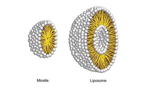 Fig. 3. Structural difference between liposomes and micelles.
- Structure: Liposomes are spherical vesicles composed of a lipid bilayer surrounded by an aqueous core. Micelles, on the other hand, are small aggregates of lipid or surfactant molecules that form spherical shapes with a hydrophobic core and a hydrophilic shell.
- Size: Liposomes are generally larger than micelles, ranging in diameter from 50 nanometers to a few micrometers. Micelles, on the other hand, are much smaller, typically in the 5 to 100 nanometer range.
- Stability: Liposomes are generally more stable than micelles. The lipid bilayer of liposomes provides a more rigid structure, making them less prone to disintegration. Micelles are smaller and less organized and more prone to rupture.
- Encapsulation capacity: Compared with micelles, liposomes have higher encapsulation capacity. The aqueous core of the liposome allows the encapsulation of both hydrophilic and lipophilic compounds. Micelles, on the other hand, are limited to encapsulating mostly lipophilic compounds within their hydrophobic cores.
- Drug Delivery: Liposomes are widely used as drug delivery systems due to their ability to encapsulate drugs and prevent drug degradation. They can also be modified to target specific tissues or cells. Micelles, although less commonly used for drug delivery, can also be used to solubilize hydrophobic drugs and enhance their bioavailability.
- Biocompatibility: Liposomes and micelles are generally considered biocompatible and have low toxicity. However, liposomes more closely resemble natural cell membranes, making them highly biocompatible and less likely to cause adverse effects.
Reference
- Duncke, A. et al. Influence of cation volume on liquid crystals of emulsified crude oil systems. 2017.
-
Lipid Nanoparticles for Small Molecule Delivery
-
Nanoparticle-based Drug Delivery Systems: Review and Current Status
-
Cholesterol: Definition, Structure, Synthesis, Types and Functions
-
Heterobifunctional PEG Linkers for Controlled Stepwise Bioconjugation
-
PEG Click Chemistry Guide: Reagents, Reactions, and Applications
-
Vaccines: Definition, History, Ingredients, Types and Mechanism of Action