Capillary Electrophoresis (CE) Technique
Capillary electrophoresis (CE) is a powerful technique that uses very narrow-bore capillaries, typically 50 µm internal diameter and 300 µm external diameter, for the high-resolution separation of large and small biomolecules such as peptides and proteins. The basic components of CE system are schematically shown in Fig. 1, including a high voltage power supply, a capillary, electrodes, two buffer reservoirs, and detector.
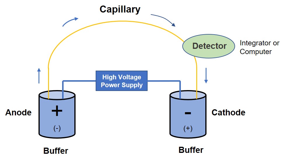 Fig. 1 Schematic diagram of capillary electrophoresis (CE) system.
How Does It Work?
The heart of the CE system is the capillary where separation takes place. In the electrophoresis process, both ends of the capillaries are inserted into the electrode solution, the buffer and analyte molecules can pass through the capillary only under the action of high voltage. In order to maintain the charge balance, the positive ions in the solution are adsorbed to the quartz surface to form a double electron layer. Under the action of high voltage, this layer will move toward the negative electrode, thus driving the solution in the capillary flow to the negative electrode, which is called electroosmosis flow.
Due to the large surface area to volume ratio of the capillary and the high voltage applied during electrophoresis, the electroosmotic flow rate is usually 5-7 times faster than the electrophoresis speed. Therefore, the use of electroosmotic flow in capillary electrophoresis (CE) can produce differential migration of positive, negative ions and neutral molecules in one direction together, realizing the separation of positive and negative ions in one operation.
Different Modes of CE
In CE, the sample is introduced by immersing the end of the capillary into a sample vial and applying pressure, vacuum or voltage. We can provide our customers with different modes of CE separation by using different types of capillary and electrolytes, including not limited to:
- Capillary Zone Electrophoresis (CZE)
- Capillary Gel Electrophoresis (CGE)
- Capillary Isoelectric Focusing (CIEF)
- Micellar Electrokinetic Capillary Chromatography (MECC)
The most basic and commonly used mode of CE for analysis of charged solutes. The separation mechanism is based on differences in the charge-to-mass ratio of the analytes.
CGE is electrophoresis in which the gel is moved into a capillary as a support. The gel is porous and acts like a molecular sieve that allows analytes can be separated according to molecular size. CGE can reduce the diffusion of analytes, and it can reach the highest column efficiency in CE.
CIEF is electrophoresis that takes place in the capillary. Under the action of high voltage, a pH gradient is established inside the capillary, and the proteins will migrate until reach their respective isoelectric points (pI), thus forming obvious zones.
In MECC, some ionic surfactants (such as sodium lauryl sulfate) are added to the buffer at concentrations that can form micelles. The solute will be partitioned between the water phase and the micellar phase. Neutral particles are separated due to their own hydrophobicity and the differences in the partitioning between the two phases. MECC enables CE to be a useful tool for separating neutral substances.
Analytes can be detected using one of several possible detection methods: UV-Vis, fluorescence, mass spectrometry, electrochemical detection and laser-induced fluorescence detection.
What Can CE Be Used for?
- CE has proved to be a powerful technique for the high-resolution separation of biomolecules such as peptides, proteins, enzymes and etc.
- Single CE or CE in combination with multiple separation techniques, taking advantage of differences in charge density, size, shape, and surface properties, can also be used for separating the PEG–protein conjugates, such PEGylated ribonuclease and lysozyme molecules, both in terms of degree of PEGylation and position.
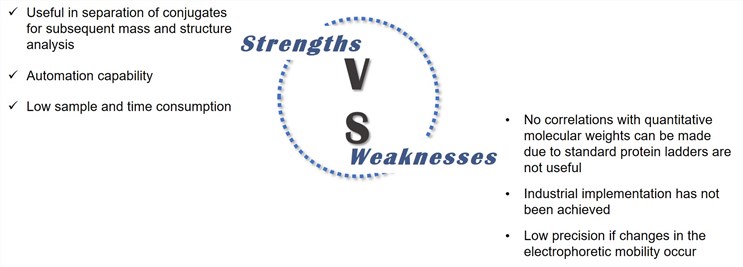
Strengths & Weaknesses of CE
References
- Payne R W, Murphy B M, et al. Product development issues for PEGylated proteins. Pharmaceutical Development and Technology, 2011, 16(5): 423-440.
- Mayolo‐Deloisa K, González‐Valdez J, et al. Current advances in the non‐chromatographic fractionation and characterization of PEGylated proteins. Journal of Chemical Technology & Biotechnology, 2011, 86(1): 18-25.